To prepare this display, a photograph of metaphase chromosomes (dyads) was cut into pieces and the individual images assembled in homologous pairs. (Now, with computer imaging, the assembly process can be done electronically.)
The staining process used here (trypsin-giemsa) reveals several hundred distinct G bands. The pattern is diagnostic for each of the chromosomes, and this permits the positive identification of chromosomes that are otherwise similar in size and shape.
The G bands also serve as landmarks for describing the location of gene loci.
| Link to an example. |
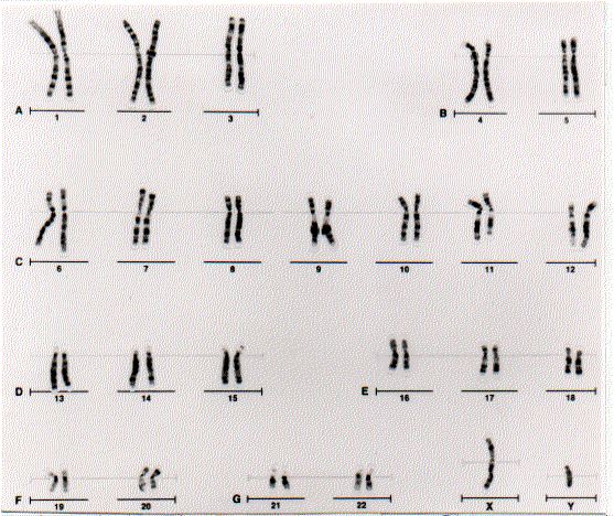
This karyotype was kindly provided by Chih-Lin Hsieh, Molecular & Clinical Cytogenetics Laboratory, Stanford University Medical Center.
| Welcome&Next Search |