
 The fluorescence-activated cell sorter is a machine that can rapidly separate the cells in a suspension on the basis of size and the color of their fluorescence.
It works like this:
This apparatus can sort as many as 300,000 cells per minute.
The cells are not damaged by the process. In fact, because the machine can be set to ignore droplets containing dead cells, the percent viability of the sorted cells can be higher than that in the original suspension.
| External Link |
| Click on this link to view an animation that shows the process far better than the static diagram here can. |
| Please let me know by e-mail if you find a broken link in my pages.) |
Often the experimenter is not interested in keeping the cells but simply in analyzing the distribution of cell size and/or surface molecules on the cells in the suspension. The signals from the detectors can generate these sorts of data, a procedure known as flow cytometry.
The data are analyzed by computer and can be plotted in several ways.
The more elaborate flow cytometers can use two different lasers to detect as many as three different colors on the cells in the suspension.
Here are some examples (all courtesy of Becton Dickinson Immunocytometry Systems).
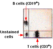
| Counting T and B cells by flow cytometry. A sample of normal human blood was treated with a fluorescent monoclonal antibody specific for the T cell surface antigen CD3 and a monoclonal antibody conjugated to a different fluorescent dye and specific for the B cell surface antigen designated CD19. Fluorescence intensity (logarithmic) is plotted on the x axis; cell number on the y axis. Note that the number of B cells is substantially less than that of T cells. The right-hand panel shows another way of plotting the data. | | |
| Counting T-cell subsets in normal human blood. Fluorescent monoclonal antibodies were directed against the CD4 molecule (left) and CD8 molecule (right). CD4+ T cells are responsible for several cell-mediated immune responses and giving help to B cells. CD8+ T cells are cytotoxic T lymphocytes (CTLs). The preponderance of CD4+ over CD8+ cells shown here is typical of healthy humans. In AIDS patients, this ratio becomes reversed and the CD4+ subset may eventually disappear. | 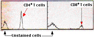 |
| Welcome&Next Search |