Gel Blotting
Gel blotting is a technique for visualizing a particular subset of macromolecules — proteins, or fragments of DNA or RNA — initially present in a complex mixture.
The steps:
- Separate the molecules by electrophoresis. This is done in a gel which allows the molecules to migrate under the influence of the electric field.
- "Blot" them with a nitrocellulose filter. For unknown reasons, the molecules stick tightly to the filter and will retain their relative positions when flooded with fluid at the next step.
- Bathe the filter with a solution containing a "probe": a molecule that
- will combine specifically with the target molecules; that is, the one(s) you are looking for
- carries a mean of visualization, e.g. a radioactive or fluorescent marker.
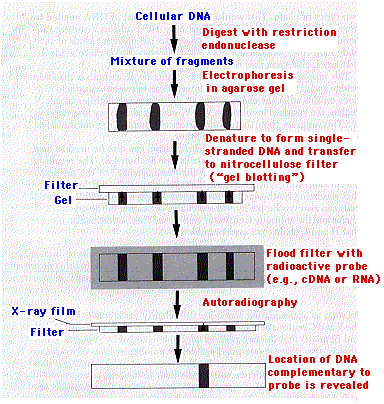
 The diagram illustrates the procedure for detecting DNA fragments containing a particular sequence.
- DNA is extracted from the cell.
- The DNA is partially digested by a restriction endonuclease.
- The resulting DNA fragments are separated by electrophoresis and then denatured to form single-stranded molecules (ssDNA).
- Without altering their positions, the separated bands of ssDNA are transferred to a nitrocellulose filter and exposed to radiolabeled cDNA or RNA.
- If the probe detects complementary DNA sequences, it will bind to them.
- The presence of the probe in a particular band is revealed by autoradiography.
This procedure was developed by E. M. Southern and the finished product is called a "Southern blot".
The same basic procedure can also be used to separate and visualize RNA molecules and protein molecules. As a humorous extension of the term "Southern blot", these have been dubbed "Northern" and "Western" blots respectively.
| Type of Blot |
Molecules separated by electrophoresis |
Probe |
| Southern |
ssDNA |
cDNA or RNA |
| Northern |
denatured RNA |
RNA or cDNA |
| Western |
Protein |
Antibodies |
11 August 2012
 The diagram illustrates the procedure for detecting DNA fragments containing a particular sequence.