The rate at which cellular respiration takes place (and hence oxygen is consumed) varies with the general state of activity of the body. Vigorous exercise may increase by 20 to 25 times the demands of the tissues for oxygen. This increased demand is met by increasing the rate and depth of breathing.

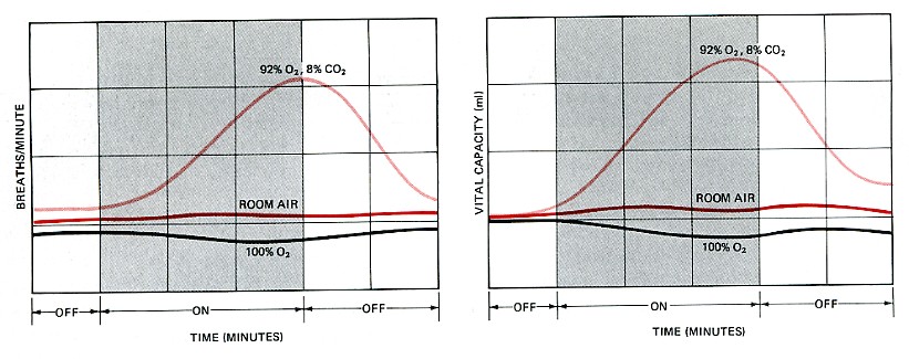
It would be reasonable to assume that lack of sufficient oxygen triggers this response. It is quite easy to show, however, that oxygen deprivation plays only a very minor role in regulating the rate at which ventilation of the lungs occurs. The figure shows an experimental setup (a modification of a device invented by my grandfather, Francis Gano Benedict) with which a human subject can inspire various precise gas mixtures. While the subject is inhaling the gas mixture, the rate and depth of breathing can be recorded.

So it is the concentration of carbon dioxide that plays the major part in governing the rate and depth of breathing.
Other experiments have shown that carbon dioxide achieves its effect through its action on the medulla oblongata. When blood passes through the alveoli, its CO2 content becomes the same as that of the alveolar air. Moments later, the blood reaches the medulla, which contains cells* that are very sensitive to the pH of the blood. As the CO2 content rises above normal levels, the pH drops [CO2 + H2O → HCO3− + H+], and the medulla oblongata responds by increasing the number and rate of nerve impulses that control the action of the intercostal muscles and diaphragm. This produces an increase in the rate of lung ventilation, which quickly brings the CO2 concentration of the alveolar air, and then of the blood, back to normal levels.
*Recent experiments in the rat (reported by Gourine, A. V., et al., in Science 30 July 2010) show that astrocytes in the medulla respond to a drop in pH by releasing ATP, which provides the stimulus for the generation of action potentials in nearby neurons.
Ventilation of the lungs is also controlled locally. The smooth muscle walls of the bronchioles are very sensitive to the concentration of CO2. A rising level causes the bronchioles to dilate. This provides less resistance in the airways and thus increases the uptake of oxygen at the same time that the increased amount of CO2 are exhaled.
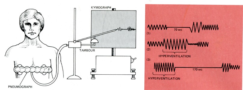
Rate and depth of breathing is, of course, also under conscious control, but this is limited. You can hold your breath for a limited period of time, but eventually the CO2 concentration in the blood reaching the medulla oblongata becomes so high that the medulla forces a breath.

This figure shows a setup with which this type of response can be studied. Movements of the subject's chest are detected by a hollow bellows (the pneumograph) strapped around the chest. Expansion and contraction of the bellows causes decreases and increases in the air within. These pressure changes are transmitted to a recording stylus, which writes on a slowly revolving drum (the kymograph).
It may seem curious that the rate at which one breathes and thus supplies oxygen to the body is controlled by carbon dioxide rather than oxygen. But cellular respiration produces CO2 as fast as oxygen is consumed, so the CO2 given off by active muscles triggers increased ventilation of the lungs and thus automatically supplies additional oxygen.
While CO2 is the major stimulus for controlling breathing, the carotid body in the carotid arteries does have receptors that respond to a drop in oxygen. Their activation is important in situations (e.g., at high altitude in the unpressurized cabin of an aircraft) where oxygen supply is inadequate, but there has been no increase in the production of CO2. People who live at high altitudes, e.g., in the Andes, have enlarged carotid bodies.| Welcome&Next Search |