
Cancer of the lung is the leading cause of death from cancer in all males and in females between the ages of 15 and 64.
Overall, the rate of new cases continues to grow even though the role of cigarette smoke in causing lung cancer is well-established.
 |
 |
| Link to discussion of the evidence linking cigarette smoking to lung cancer. |
Most cancers in the lung are carcinomas - cancers of epithelial cells.
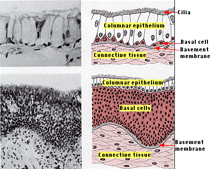
The top pair of images shows the normal epithelium of the bronchi. This photomicrograph is greatly enlarged.
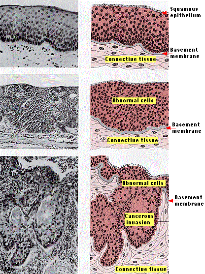
The next three pairs of images show three successive stages of abnormal changes in the bronchial epithelium commonly found in smokers. The drawings show the cellular organization occurring at each stage.
These stages show a well-defined sequence of cellular changes:
At later stages, however, the cells have lost the ability to stop dividing; they are now cancerous. But as long as they do not penetrate the basement membrane, they do not metastasize; that is, do not spread in blood and lymph to other parts of the body.
This stage is called carcinoma in situ.If the lesion is removed at this stage, the patient is cured.
The final pair of images shows the cancerous lesion having broken through the basement membrane (also known as the basal laminar). This is now a full-blown case of lung cancer.
(All the photomicrographs courtesy of Oscar Auerbach, M.D.)It is the metastasis of these cells to other parts of the body that will kill the patient (most within a year of being diagnosed).
Other carcinomas such asGenetic analysis reveals that the structural changes seen at each step reflect an accumulation of mutations in
Although tumor-suppressor genes are recessive, mutations in one may be accompanied by a loss of the other allele (called loss-of-heterozygosity).
These cells are all members of a clone — the descendants of a single original cell. But as a descendant cell of the clone acquires another mutation, the growth advantage it imparts enables that cell to proliferate and overgrow its less well-endowed cousins.
The 14 January 2010 issue of Nature contains the most detailed analysis yet of the genetic changes in lung cancer (Pleasance, E. D., et al., Nature: 463:184, 2010). These workers (there were 40 authors listed) sequenced the complete genome of a lung cancer and compared it with the sequence of a noncancerous tissue from the same patient. They found a total of 22,910 somatic mutations in the tumor DNA that were not in the normal genome. The vast majority of these were "silent" and thus seemed to be "passengers" not "drivers" of the malignant state. However, 134 mutations were in protein-coding exons including some already-familiar culprits like p53, and RB. And some mutations that did not alter protein structure may well have occurred in control regions, i.e. promoters and enhancers, and thus be drivers as well.
Calculating from the average smoking history of lung cancer patients, these workers estimated that the single cell line that ultimately grew into the cancer suffered another mutation with every 15 cigarettes smoked.
The overall death rate among former smokers is inversely proportional to the time elapsed since they gave up the habit. Sequencing the genome of individual lung cells of former smokers reveals a growing population of healthy cells and a diminishing population of cells carrying the high load of mutations found in lung cancer.
| Link to the genetic alterations characteristic of the development of colon cancer. |
| Welcome&Next Search |